


Research

 Research topics

Innovation
Grant Support
 Main Research Lines

Hadron Therapy
 Hadron therapy is a cancer treatment technique that employs massive charged particles, generally protons or Carbon ions. This treatment is advantageous over conventional X-ray radiotherapy, since it allows concentration of the radiation dose in smaller volume (the maximal dose is deposited at a point known as the Bragg peak) and reduction of the overdose absorbed by the patient's healthy tissue. In order to exploit the beneficial features of hadron therapy, it is necessary rely on monitoring systems to verify the correct dose deposition during treatment. Secondary particles emitted during irradiation can be employed to this end.
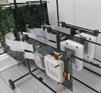
Compton telescope. The IRIS group works on on the development of a Compton telescope based on the novel technology of LaBr3 crystals coupled to SiPMs for the monitoring of the radiation dose delivered to the patient. MACACO (Medical Applications CompAct COmpton camera) is a three-layer Compton telescope that we have developed with its associated readout electronics and image reconstruction code. The first prototype version was developed and successfully tested in the laboratory, where images with gamma-rays at therapeutic energies were obtained. It was also tested on-beam at accelerator facilities, where Bragg peak shifts were detected with 150 MeV protons. The prototype improvent with new detectors leading to an enhanced resolution and advanced image reconstruction codes has allowed imaging more complex sources. Distributions of photons emitted during irradiation of a PMMA target with a proton beam have also been imaged.
Coaxial prompt gamma-ray monitoring. Several gamma-ray imaging prototypes are being developed for range verification in proton therapy, typically comprising between ten and hundred electronic channels, and are undergoing first clinical studies with promising results. To facilitate the translation of this technology to the routine clinical workflow, we have developed a novel prototype that minimizes cost and required space by using a single detector channel withstanding extreme count rates of up to 10 million gamma-rays per second using pile-up recovery techniques. When operated in a coaxial geometry (downstream from the patient, aligned with the beam axis), a proton range over or undershoot is seen as an increase or decrease in the detection count rate. Its sensitivity has been investigated with standard Monte Carlo simulations. A fast GPU Monte Carlo simulation of the detector using SYCL is under development, including a visual interface with 3D Slicer (SlicerRT) is under development for early feedback to the physician. Further simulations of the photodetector and voltage divider circuit have been carried out.
Positron Emission Tomography
The IRIS group works on the development of innovative Positron Emission Tomography PET systems.
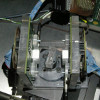 PETETE. The IRIS group has developed a PET system for small animals with continuous crystals and SiPMs instead of the pixellated (segmented) detectors generally employed by conventional systems. Continuous cristals can provide high efficiency together with high spatial resolution, but the determination of the interaction position of the 511 keV photons in the crystal becomes challenging. The group has adopted a method for 3D (including depth of interaction) determination of the interaction position in the crystal that is giving excellent results, with a spatial resolution of 0.7 mm FWHM achieved.
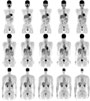
We have assembled a first prototype (PETETE- PET Exploring Techniques for performance Enhancement) consisting of two detector heads rotating around the source. PETETE is a high resolution PET for small animals. Each head is composed of a continuous LYSO crystal coupled to a 64-channel monolithic silicon photomultiplier (SiPM) matrix of 12 mm x 12 mm size. The matrix has 8 x 8 SiPM elements in a common substrate, with 1.5 mm pitch. We have developed an image reconstruction code that profits from the benefits of continuous crystals and we have reconstructed images of a Na-22 sources in different positions of the field-of-view and of a phantom filled with FDG.
PETETE. The IRIS group has developed a PET system for small animals with continuous crystals and SiPMs instead of the pixellated (segmented) detectors generally employed by conventional systems. Continuous cristals can provide high efficiency together with high spatial resolution, but the determination of the interaction position of the 511 keV photons in the crystal becomes challenging. The group has adopted a method for 3D (including depth of interaction) determination of the interaction position in the crystal that is giving excellent results, with a spatial resolution of 0.7 mm FWHM achieved.
We have assembled a first prototype (PETETE- PET Exploring Techniques for performance Enhancement) consisting of two detector heads rotating around the source. PETETE is a high resolution PET for small animals. Each head is composed of a continuous LYSO crystal coupled to a 64-channel monolithic silicon photomultiplier (SiPM) matrix of 12 mm x 12 mm size. The matrix has 8 x 8 SiPM elements in a common substrate, with 1.5 mm pitch. We have developed an image reconstruction code that profits from the benefits of continuous crystals and we have reconstructed images of a Na-22 sources in different positions of the field-of-view and of a phantom filled with FDG.
Total-body PET spatial resolution improvement. One current line of research in nuclear medicine is the study of a total-body scanner, the development of which would represent an improvement on the current features of PET scannersin terms of sensitivity and field-of-view. Currently, the typical spatial resolution of the reconstructed PET images is around 5 mm. The use of PET probes would improve spatial resolution without compromising the scanner sensitivity. A study of the suitability of a PET probe for this task, based on scintillation crystals and silicon photomultipliers (SiPM), is being carried out. The functioning of the probe will be tested in a Preclinical Super Argus PET/CT scanner for small animals.
Research topics
Research data associated with published articles of the group are available to any external researcher upon reasonable request to our e-mail address iris at ific dot uv dot es.
Detector development
The group works on the development of instrumentation for hadron therapy monitoring and for PET. We are experts in PET and Compton cameras and we employ the newest technologies and innovative techniques beyond the state of the art. Our developments include detectors, electronics, data acquisition codes and data analysis algorithms. Our work is currently based on continuous crystals coupled to SiPMs and diffeent types of silicon detectors.
Image Reconstruction in PET
 The aim of image reconstruction in PET is to find the radioisotope distribution that originated the data measured by the scanner,
what mathematically is call an "inverse problem". Although there is no exact solution, there are many reconstruction methods
that provide an approximate solution; to this end mathematical algorithms and powerful computers are required.
The aim of image reconstruction in PET is to find the radioisotope distribution that originated the data measured by the scanner,
what mathematically is call an "inverse problem". Although there is no exact solution, there are many reconstruction methods
that provide an approximate solution; to this end mathematical algorithms and powerful computers are required.
Our groups investigates, mainly, in the application of iterative statistical algorithms combined with detailed physical models of the image formation process.
Physical models for phenomena involved in image formation and degradation processes
 The iterative algorithms require the inclusion of a model describing the response of a scanner to the presence of radiation
(system matrix). The accuracy and precision of the model will have a great impact in the final image. In order to obtain
high resolution and quality images, it will be necessary to have detailed models that include a description of those phenomena
that take part in the image formation process, as the gamma ray interactions with the detectors.
The image quality can be further improved if, in addition, it is included in the model a description of phenomena that
degrade the image, as for instance, gamma ray interactions in the subject.
The iterative algorithms require the inclusion of a model describing the response of a scanner to the presence of radiation
(system matrix). The accuracy and precision of the model will have a great impact in the final image. In order to obtain
high resolution and quality images, it will be necessary to have detailed models that include a description of those phenomena
that take part in the image formation process, as the gamma ray interactions with the detectors.
The image quality can be further improved if, in addition, it is included in the model a description of phenomena that
degrade the image, as for instance, gamma ray interactions in the subject.
Compensation of image degradation phenomena
 There are several physical effects, inherent to PET, that contribute to degrade the image; for correcting or compensating
these effects it is necessary to have models that describe them properly. Some models can be include in the system matrix,
like the positron range and the acollinearity of the emitted photons, or the attenuation suffered by the photon inside the
subject. Other effects are treated in the measure space, like photon scatter in the subject, or ranom coincidences. In most
cases models are approximated.
There are several physical effects, inherent to PET, that contribute to degrade the image; for correcting or compensating
these effects it is necessary to have models that describe them properly. Some models can be include in the system matrix,
like the positron range and the acollinearity of the emitted photons, or the attenuation suffered by the photon inside the
subject. Other effects are treated in the measure space, like photon scatter in the subject, or ranom coincidences. In most
cases models are approximated.
Our group is interested in improving the traditional models by a more precise formulation of the problem, like in the case of random coincidences. We also investigate the contribution of other sources of image degradation that usually are neglected in conventional PET. We study the way to compensate those effects with the use of statistical descriptions. Our final goal is to improve the quality and resolution of the final image.
Monte-Carlo simulations for PET
 Monte-Carlo simulations are a useful tool in many fields of science and technology.
In physics, they are employed mainly in Nuclear and Particle Physics; in Medical Physics they are used mostly in radiotherapy
planification for dose computation.
In Medical Image, Monte-Carlo simulation techniques have multiple uses: they make possible to optimize the design of new
prototypes and evaluate their performance, understand and analize the underlying phenomena in the image generation, disentangle
effects that in real situations appear together, or generate data to be used for validating novel recontruction algorithms.
Monte-Carlo simulations are a useful tool in many fields of science and technology.
In physics, they are employed mainly in Nuclear and Particle Physics; in Medical Physics they are used mostly in radiotherapy
planification for dose computation.
In Medical Image, Monte-Carlo simulation techniques have multiple uses: they make possible to optimize the design of new
prototypes and evaluate their performance, understand and analize the underlying phenomena in the image generation, disentangle
effects that in real situations appear together, or generate data to be used for validating novel recontruction algorithms.
Nowadays, there are several simulation packages optimized for their use in Medical Image, like SimSet or GATE, although, in same cases, other packages devised for High Energy Physics, like Geant4, can be more useful. Sometimes, it can be desirable to write from scratch the code for the simulation.
In IRIS we use Geant4, GATE and private codes to develope and validate models of relevant physical phenomena in PET (like Compton scatter or random coincidences), with the aim of studying the impact of the above mentioned phenomena in the final image, or for forseing the performance of novel designs.
Innovation
The IRIS group also carries out innovation and technology transfer activities, participating in R+D contracts with companies, technology transfer projects and at IFIC's UCIE activities with two projects. The group has patented a data acquisition system that is currently commercialized by Alibava Systems S.L.
Grant Support
AIDER: Advanced Imaging DEtector for targeted Radionuclide therapy (GA. num 101165088)
EU HORIZON-EURATOM 2023.
Participants: IFIC (Coordinator), UCBL Lyon, Universität Zu Lübeck, Politecnico di Milano, HUIP La Fe, Hospital Léon Bérard Lyon, DAMAVAN Imaging.
Duration: 1/9/2025 - 31/8/2029.
Funding: 3 499 196.25 EUR. IFIC: 712 792.50 EUR.
Coordinator and PI IFIC:
Dra. Gabriela Llosá
Conference organization: ANIMMA conference (CIAORG/2024/073)
Duration: 1/9/2025 - 31/8/2029.
Funding: 9000 EUR.
Coordinator and PI IFIC:
Dra. Gabriela Llosá
FARO: Feasibility of Adaptive Surface-Guided Radiation Oncology (AP2024VLC-01)
VLC Biomed programme. Preparatory actions UV / IIS La Fe.
Participants: IFIC (Universidad de Valencia / CSIC) and HUiP La Fe.
Duration: 4/2/2025 - 4/2/2026.
Funding: 7500 EUR.
Principal investigators:
Dr. Ana Ros García (UV) and Dr. Montserrat Carles Fariña (HUiP La Fe)
PTCOG Project Funding 2024 (Physics)
PTCOG.
Duración: 01/07/24-30/06/25
Financiación: 9.000 EUR.
P.I.: Dr. Fernando Hueso-González
El tragaluz cuántico (FCT-23-18895)
FECYT.
Duración: 01/07/24-30/06/25
Financiación: 27.000 EUR.
P.I at UPV: Jaime Riera.
P.I. at IFIC:
Dr. Fernando Hueso-González
IMMPRINT-Integrated molecular Imaging for Personalized Biomarker-based Breast Cancer Characterization and Treatment (EURATOM research and innovation program - 101061037 grant agreement)
European's Union.
Duración: 01/04/24-31/03/27
Financiación: 295.155 EUR.
P.I. at IFIC:
Dra. Gabriela Llosá
HIgh-COunt RatE photon detection with scintillators coupled to photomultiplier tubes and fast digitizers (RADNEXT TA09-275)
European Union's Horizon 2020 (grant agreement No 101008126).
Duración: 01/07/24-30/06/25
Financiación: 10.500 EUR.
P.I.:
Dr. Fernando Hueso-González
MIDAS - Medical imaging for Improved Diagnostics And Treatments (PID2022-143246OB-I00)
Ministerio de Ciencia, Innovación y Universidades.
Participants: IFIC (Universidad de Valencia / CSIC), Univ. of Lübeck, Univ. of Sherbrooke, Univ. of Lyon.
Duración: 01/09/2023-31/08/2026
Financiación: 98.371 EUR.
Investigadoras principales:
Dra. Gabriela Llosá
ICOR - Imagen Compton para terapia con radionúclidos (ASFAE/2022/019)
MCIU, through Plan de Recuperación, Transformación y Resiliencia (PRTR-C17.I01), with funding from the European Union NextGenerationEU and Generalitat Valenciana (GVANEXT).
Duración: 1/9/2022-31/12/2025
Financiación: 299.920 EUR.
Investigadoras principales:
Dra. Gabriela Llosá y Dra. Irene Torres Espallardo
VALID - Valorization of new detectors for medical imaging (PDC2021-121839-I00)
Ministerio de Ciencia e Innovación.
Participants: IFIC (Universidad de Valencia / CSIC), Centro de protonterapia Quirónsalud.
Duration: 1/12/21-30/11/23.
Funding: 115000 EUR.
Principal investigator:
Dr. Gabriela Llosá
VALMONT- Valorization of a hadron therapy monitoring system (INNVA1/2021/37)
Agencia Valenciana de Innovación (AVI). Generalitat Valenciana.
Participants: IFIC (Universidad de Valencia / CSIC).
Duration: 1/1/2021 – 30/09/2023.
Funding: 337926.35 EUR.
Principal investigator:
Dr. Gabriela Llosá
MONDO- Monitoring and dosimetry in hadron therapy (PID2019-110657RB-I00)
Ministerio de Ciencia e Innovación.
Participants: IFIC (Universidad de Valencia / CSIC).
Duration: 1/6/2020 - 31/05/2023.
Funding: 98010 EUR.
Principal investigator:
Dr. Gabriela Llosá
New-TIM: New technologies in medical imaging (PI2020-16)
VLC Biomed programme. Innovation projects UV / IIS La Fe.
Participants: IFIC (Universidad de Valencia / CSIC) and HUiP La Fe.
Duration: 1/4/2021 - 31/03/2022.
Funding: 19000 EUR.
Principal investigators:
Dr. Gabriela Llosá (UV) and Irene Torres (HUiP La Fe)
Novel cost-effective proton range verification based on coaxial prompt gamma-ray monitoring (CDEIGENT/2019/011)
Generalitat Valenciana. Conselleria de Educación, Investigación, Cultura y Deporte.
Participants: IFIC (Universidad de Valencia / CSIC).
Duration: 1/6/2020 - 31/05/2026.
Funding: 357250 EUR.
Principal investigator:
Dr. Fernando Hueso-González
Implementación y demostración de la funcionalidad de una nueva sonda para el aumento de la resolución espacial en tomografía por emisión de positrones (PET) (SEJI/2019/014)
Generalitat Valenciana. Conselleria de Educación, Investigación, Cultura y Deporte.
Participants: IFIC (Universidad de Valencia / CSIC).
Duration: 1/9/2019 - 31/12/2021.
Funding: 159800 EUR.
Principal investigator:
Dr. Ana Ros García
INCONI: Intercomparison of Compton cameras for medical applications. (2018FR0032)
CSIC PICS 2019.
Participants: IFIC (Universidad de Valencia / CSIC). CNRS, Lyon, France.
Duration: 1/1/2019 - 31/12/2021.
Funding: 10000 EUR.
Principal investigator:
Dr. Gabriela Llosá
Estimación de dosis en terapia hadrónica (AICO/2019/070)
Generalitat Valenciana. Conselleria de Educación, Investigación, Cultura y Deporte.
Participants: IFIC (Universidad de Valencia / CSIC). Universidad de Alicante. Universidad de Murcia.
Duration: 1/1/2019 - 31/12/2020.
Funding: 39927 EUR.
Principal investigator:
Dr. Carlos Lacasta
COBRA- Compton Based Research Applications (FPA2017-85611-R)
Ministerio de Economía, Industria y Competitividad.
Participants: IFIC (Universidad de Valencia / CSIC).
Duration: 1/1/2018 - 31/12/2020.
Funding: 60000 EUR.
Principal investigator:
Dr. Gabriela Llosá and Prof. José Bernabéu
ENSAR2 - MediNet (grant agreement num 654002)
European Commission. H2020.
Participants: 30 European institutions.
Duration: 1/3/2016 - 28/2/2020.
Funding: 10 MEUR.
Principal investigator:
Dr. Peter Thirolf
Multidetector DAQ system (UV-INV-PROVAL17-720859)
Universitat de València.
Participants: IFIC (Universidad de Valencia / CSIC).
Duration: 7/9/2018 - 6/9/2019.
Funding: 44873.88 EUR.
Principal investigator:
Dra. Gabriela Llosá
Detectores para aplicaciones médicas (FPA2014-53599-R)
Ministerio de Economía y Competitividad.
Participants: IFIC (Universidad de Valencia / CSIC), HUiP La Fe.
Duration: 1/1/2015 - 31/12/2017.
Funding: 30250 EUR.
Principal investigator:
Dra. Gabriela Llosá
Alineamiento Temporal de sonda y escáneres comerciales (18_ATIME_LLOSA_BELLO_2014)
Participants: IFIC (Universidad de Valencia / CSIC), Hospital Universitario y Politécnico La Fe.
Duration: 1 May 2015 - 31 Dec 2015.
Principal investigator:
Dra. Gabriela Llosá y Dra. Pilar Bello
Development of detectors for compton telescopes (GV/2013/133)
Generalitat Valenciana.
Participants: IFIC (Universidad de Valencia / CSIC).
Principal investigator:
Dra. Gabriela Llosá
High resolution and efficiency detectors for medical applications. (UV-INV-PRECOMP12-80755)
Universitat de València.
Participants: IFIC (Universidad de Valencia / CSIC).
Principal investigator:
Dra. Gabriela Llosá
ASPID: Application of Silicon Photomultipliers to Imaging Detectors.
EU-FP7. European Reintegration Grant. GA num 239362.
Participants: CSIC.
Fellow: Dra. Gabriela Llosá
Scientist in Charge:
Dr. Carlos Lacasta
Image quality and quantification in Positron Emission Tomography (FPA2010-14891)
Plan Nacional de I-D-i, Spanish Ministry of Science and Innovation.
Participants: IFIC (Universidad de Valencia / CSIC), RWTH Aachen (Germany).
Principal investigator:
Dr. Magdalena Rafecas
Compton telescope for monitorization in hadron therapy (FIS2011-14585-E)
Plan Nacional de I-D-i, Spanish Ministry of Science and Innovation.
Participants: IFIC (Universidad de Valencia / CSIC).
Principal investigator:
Dr. Magdalena Rafecas
Innovative Software for Advanced PET Imaging (INSPET) (PIEFGA2009237620)
European Comission.
Participants: IFIC (Universidad de Valencia / CSIC).
Principal investigator:
Dr. Magdalena Rafecas
European NoVel Imaging Systems for ION therapy (ENVISION)
European Comission.
Participants: CERN (Suiza), TU Dresden (Alemania), Medizinische Universitaet Wien (Austria), Universite Claude Bernard Lyon 1 (Francia), Oxford University (G.B.), Siemens AG (Alemania), Ion Beam Applications SA (Belgica), INFN (Italia), Fondazione per adroterapia oncologica (Italia), Universitaetsklinikum Heidelberg (Alemania), GSI (Alemania), Maastro clinic (Holanda), CNRS (Francia), CSIC, Universiteit Gent, Politecnico di Milano (Italia), Universidad de Castilla - La Mancha.
Principal investigator:
Prof. Manjit Dosanjh (CERN)
Grid y E-Cencia: Análisis de Datos Atlas y Física Médica (PCI A/023372/10)
Ministerio de asuntos exteriores y Agencia Española de Cooperación Internacional para el Desarrollo (AECID).
Participants: Universitat de València with the Centre National de Reserche Scientifique et Technologique (CNRST) and Mohamed V-Agdal University (Marruecos).
Principal investigator:
Dr. Santiago González de la Hoz
AX-PET: Un nuevo concepto de escáner para tomografía por emisión de positrones con WLS-Strips y G-APDs (FPA2008-02419-E/FPA)
Spanish Ministry of Education and Science / Complementary actions.
Participants: IFIC (CSIC/Universidad de Valencia).
Principal investigator:
Dr. Magdalena Rafecas
Improving image quality in positron emission tomography
Plan Nacional de I-D-i, Spanish Ministry of Education and Science.
Participants: IFIC (Universidad de Valencia / CSIC), Université de Sherbrooke (Sherbrooke, Canada),
Technische Universität München (Munich, Germany).
Principal investigator:
Dr. Magdalena Rafecas
Tomografía por emisión de positrones para pequeños animales: Coincidencias accidentales y dispersión Compton entre cristales
"Acciones Complementarias Internacionales", Spanish Ministry of Education and Science.
Participants: IFIC (Universidad de Valencia / CSIC), Université de Sherbrooke (Sherbrooke, Canada).
Principal investigator:
Dr. Magdalena Rafecas
Development of a small animal PET tomograph based on novel silicion photomultipliers, (HI2006-0082)
"Acciones Integradas", Spanish Ministry of Education and Science.
Participants: IFIC (Universidad de Valencia / CSIC), Universitá degli Studi di Pisa (Pisa, Italy).
Principal investigator:
Dr. Magdalena Rafecas
(Spain) / Prof. Alberto del Guerra (Italy)
MADEIRA: Minimizing activity and dose with enhanced image quality by radiopharmaceutical administration
EUROATOM, European Comission.
Participants: Helmholtz-Insitut Mnchen (Germany), Lund University (Sweden), Jozef Stefan Institute (Slovenia), IFIC (Spain),
Scivis wissenschaftliche Bildverarbeitung (Germany), Universitá degli Studi di Milano (Italy), The University of Michigan (USA).
Principal investigator:
Prof. Christoph Hoeschen,
(Helmholtz-Institut München).
Toward sensitivity improvement of positron emission tomography imaging by including low energy photons in images
Strategic Plan of the Department of Electrical Engineering, Université de Sherbrooke (Canada).
Participants: Université de Sherbrooke, IFIC (Universidad de Valencia / CSIC).
Principal investigator: Prof. Réjean Fontaine (Université de Sherbrooke).
PARTNER: Particle Training Network for European Radiotherapy
Marie Curie Initial Training Networks (ITN), European Comission.
Participants: CERN, CNAO (Italy), GSI (Germany), Heidelberg Ion
Therapy Inc. (Germany), Karolinska Institutet (Sweden),
University of Surrey (UK), TERA Foundation (Italy),
Ion Beam Applications, RaySearch
Laboratories, Siemens Medical, ETOILE Project (France), IFIC (Spain), Med-Austron (Austria).
Principal investigator: Prof. Manjit Dosanjh (CERN, Switzerland)
