

Research

 Research topics

Scanners
Grant Support
 Research Topics

Image Reconstruction in PET
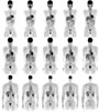 The aim of image reconstruction in PET is to find the radioisotope distribution that originated the data measured by the scanner,
what mathematically is call an "inverse problem". Although there is no exact solution, there are many reconstruction methods
that provide an approximate solution; to this end mathematical algorithms and powerfull computers are required.
The aim of image reconstruction in PET is to find the radioisotope distribution that originated the data measured by the scanner,
what mathematically is call an "inverse problem". Although there is no exact solution, there are many reconstruction methods
that provide an approximate solution; to this end mathematical algorithms and powerfull computers are required.
Our groups investigates, mainly, in the application of iterative statistical algorithms combined with detailed physical models of the image formation process.
Physical Models for phenomena involved in image formation and degradation processes
 The iterative algorithms require the inclusion of a model describing the response of a scanner to the presence of radiation
(system matrix). The accuracy and precision of the model will have a great impact in the final image. In order to obtain
high resolution and quality images, it will be necessary to have detailed models that include a description of those phenomena
that take part in the image formation process, as the gamma ray interactions with the detectors.
The image quality can be further improved if, in addition, it is included in the model a description of phenomena that
degrade the image, as for instance, gamma ray interactions in the subject.
The iterative algorithms require the inclusion of a model describing the response of a scanner to the presence of radiation
(system matrix). The accuracy and precision of the model will have a great impact in the final image. In order to obtain
high resolution and quality images, it will be necessary to have detailed models that include a description of those phenomena
that take part in the image formation process, as the gamma ray interactions with the detectors.
The image quality can be further improved if, in addition, it is included in the model a description of phenomena that
degrade the image, as for instance, gamma ray interactions in the subject.
In GFIM we use, mostly, Monte-Carlo simulatios to obtain the system matrix. Our aim is to compensate especially the effects derived form the radiation penetration through the crystals, since this phenomenon is the main cause of spatial resolution decreasing far from the the field of view center for high resolution scanners of small diameter, as those designed for small animals.
Compensation of image degradation phenomena
 There are several physical effects, inherent to PET, that contribute to degrade the image; for correcting or compensating
these effects it is necessary to have models that describe them properly. Some models can be include in the system matrix,
like the positron range and the acollinearity of the emitted photons, or the attenuation suffered by the photon inside the
subject. Other effects are treated in the measure space, like photon scatter in the subject, or ranom coincidences. In most
cases models are approximated.
There are several physical effects, inherent to PET, that contribute to degrade the image; for correcting or compensating
these effects it is necessary to have models that describe them properly. Some models can be include in the system matrix,
like the positron range and the acollinearity of the emitted photons, or the attenuation suffered by the photon inside the
subject. Other effects are treated in the measure space, like photon scatter in the subject, or ranom coincidences. In most
cases models are approximated.
Our group is interested in improving the traditional models by a more precise formulation of the problem, like in the case of random coincidences. We also investigate the contribution of other sources of image degradation that usually are neglected in conventional PET. We study the way to compensate those effects with the use of statistical descriptions. Our final goal is to improve the quality and resolution of the final image.
Monte-Carlo simulations for PET
 Monte-Carlo simulations are a useful tool in many fields of science and technology.
In physics, they are employed mainly in Nuclear and Particle Physics; in Medical Physics they are used mostly in radiotherapy
planification for dose computation.
In Medical Image, Monte-Carlo simulation techniques have multiple uses: they make possible to optimize the design of new
prototypes and evaluate their performance, understand and analize the underlying phenomena in the image generation, disentangle
effects that in real situations appear together, or generate data to be used for validating novel recontruction algorithms.
Monte-Carlo simulations are a useful tool in many fields of science and technology.
In physics, they are employed mainly in Nuclear and Particle Physics; in Medical Physics they are used mostly in radiotherapy
planification for dose computation.
In Medical Image, Monte-Carlo simulation techniques have multiple uses: they make possible to optimize the design of new
prototypes and evaluate their performance, understand and analize the underlying phenomena in the image generation, disentangle
effects that in real situations appear together, or generate data to be used for validating novel recontruction algorithms.
Nowadays, there are several simulation packages optimized for their use in Medical Image, like SimSet or GATE, although, in same cases, other packages devised for High Energy Physics, like Geant4, can be more useful. Sometimes, it can be desirable to write from scratch the code for the simulation.
In GFIM we use Geant4, GATE and private codes to develope and validate models of relevant physical phenomena in PET (like Compton scatter or random coincidences), with the aim of studying the impact of the above mentioned phenomena in the final image, or for forseing the performance of novel designs.
Scanners
MADPET-II
 MADPET-II is the second PET prototype for small animals built by the
Instrumentation Group
of the
Technische Universität Mnchen. This prototype is characterized by an individual readout of its 1152 LSO crystals
through Avalanche Photodiodes (APDs), arranged in two concentric layers of 18 modules each. Another singular feature
of MADPET-II is the acquisition of "singles" events in list mode, instead of the conventional acquisition of pairs of
events detected in coincidence.
MADPET-II is the second PET prototype for small animals built by the
Instrumentation Group
of the
Technische Universität Mnchen. This prototype is characterized by an individual readout of its 1152 LSO crystals
through Avalanche Photodiodes (APDs), arranged in two concentric layers of 18 modules each. Another singular feature
of MADPET-II is the acquisition of "singles" events in list mode, instead of the conventional acquisition of pairs of
events detected in coincidence.
LabPET TM
 The new LabPET TM system is a second-generation APD-based animal PET scanner that combines avalanche photodiode (APD) technology with a highly integrated, fully digital, parallel electronic architecture.
LabPET has been
developed at the Université de Sherbrooke (Québec, Canadá, and it is now commercialized by
AMI-Gamma Medica.
The LabPET quad detector module consists of LYSO/LGSO phoswich assemblies individually coupled to reach-through APDs
Individual crystals 2x2x~10 mm3 in size are optically coupled in pair along one long side to form the phoswich detectors.
Good energy resolution and decay time difference allow for efficient crystal identification by pulse-shape discrimination.
The new LabPET TM system is a second-generation APD-based animal PET scanner that combines avalanche photodiode (APD) technology with a highly integrated, fully digital, parallel electronic architecture.
LabPET has been
developed at the Université de Sherbrooke (Québec, Canadá, and it is now commercialized by
AMI-Gamma Medica.
The LabPET quad detector module consists of LYSO/LGSO phoswich assemblies individually coupled to reach-through APDs
Individual crystals 2x2x~10 mm3 in size are optically coupled in pair along one long side to form the phoswich detectors.
Good energy resolution and decay time difference allow for efficient crystal identification by pulse-shape discrimination.
AX-PET
![]() This prototype, under construction at the moment, uses several LYSO crystal layer, axially oriented, and coupled to a APD in
Geiger mode (G-APDs), also known as Silicon Photomultipliers (Si-PMs). The z coordenate of the interaction is obtained through
"wave length shifting strips" (WLSs) arranged perpendicularly under each layer of crystals. For the WLSs readout G-APDs are
also used.
The AX-PET collaboration
takes charge of the development of this prototype. In the colaboration take part: INFN, CERN, University of Michigan, University of Ohio, University of Oslo, PSI, Instituto Superiore di Sanita, IFIC (CSIC-UV), ETH, University Hospital Geneva.
This prototype, under construction at the moment, uses several LYSO crystal layer, axially oriented, and coupled to a APD in
Geiger mode (G-APDs), also known as Silicon Photomultipliers (Si-PMs). The z coordenate of the interaction is obtained through
"wave length shifting strips" (WLSs) arranged perpendicularly under each layer of crystals. For the WLSs readout G-APDs are
also used.
The AX-PET collaboration
takes charge of the development of this prototype. In the colaboration take part: INFN, CERN, University of Michigan, University of Ohio, University of Oslo, PSI, Instituto Superiore di Sanita, IFIC (CSIC-UV), ETH, University Hospital Geneva.
Grant Support
Image quality and quantification in Positron Emission Tomography (FPA2010-14891)
Plan Nacional de I-D-i, Spanish Ministry of Science and Innovation.
Participants: IFIC (Universidad de Valencia / CSIC), RWTH Aachen (Germany).
Principal investigator:
Dr. Magdalena Rafecas
Compton telescope for monitorization in hadron therapy (FIS2011-14585-E)
Plan Nacional de I-D-i, Spanish Ministry of Science and Innovation.
Participants: IFIC (Universidad de Valencia / CSIC).
Principal investigator:
Dr. Magdalena Rafecas
Innovative Software for Advanced PET Imaging (INSPET) (PIEFGA2009237620)
European Comission.
Participants: IFIC (Universidad de Valencia / CSIC).
Principal investigator:
Dr. Magdalena Rafecas
European NoVel Imaging Systems for ION therapy (ENVISION)
European Comission.
Participants: CERN (Suiza), TU Dresden (Alemania), Medizinische Universitaet Wien (Austria), Universite Claude Bernard Lyon 1 (Francia), Oxford University (G.B.), Siemens AG (Alemania), Ion Beam Applications SA (Belgica), INFN (Italia), Fondazione per adroterapia oncologica (Italia), Universitaetsklinikum Heidelberg (Alemania), GSI (Alemania), Maastro clinic (Holanda), CNRS (Francia), CSIC, Universiteit Gent, Politecnico di Milano (Italia), Universidad de Castilla - La Mancha.
Principal investigator:
Prof. Manjit Dosanjh (CERN)
Grid y E-Cencia: Análisis de Datos Atlas y Física Médica (PCI A/023372/10)
Ministerio de asuntos exteriores y Agencia Española de Cooperación Internacional para el Desarrollo (AECID).
Participants: Universitat de València with the Centre National de Reserche Scientifique et Technologique (CNRST) and Mohamed V-Agdal University (Marruecos).
Principal investigator:
Dr. Santiago Gonzalez de la Hoz
AX-PET: Un nuevo concepto de escáner para tomografía por emisión de positrones con WLS-Strips y G-APDs (FPA2008-02419-E/FPA)
Spanish Ministry of Education and Science / Complementary actions.
Participants: IFIC (CSIC/Universidad de Valencia).
Principal investigator:
Dr. Magdalena Rafecas
Improving image quality in positron emission tomography
Plan Nacional de I-D-i, Spanish Ministry of Education and Science.
Participants: IFIC (Universidad de Valencia / CSIC), Université de Sherbrooke (Sherbrooke, Canada),
Technische Universität Mnchen (Munich, Germany).
Principal investigator:
Dr. Magdalena Rafecas
Tomografía por emisión de positrones para pequeños animales: Coincidencias accidentales y dispersión Compton entre cristales
"Acciones Complementarias Internacionales", Spanish Ministry of Education and Science.
Participants: IFIC (Universidad de Valencia / CSIC), Université de Sherbrooke (Sherbrooke, Canada).
Principal investigator:
Dr. Magdalena Rafecas
Development of a small animal PET tomograph based on novel silicion photomultipliers, (HI2006-0082)
"Acciones Integradas", Spanish Ministry of Education and Science.
Participants: IFIC (Universidad de Valencia / CSIC), Università degli Studi di Pisa (Pisa, Italy).
Principal investigator:
Dr. Magdalena Rafecas
(Spain) / Prof. Alberto del Guerra (Italy)
MADEIRA: Minimizing activity and dose with enhanced image quality by radiopharmaceutical administration
EUROATOM, European Comission.
Participants: Helmholtz-Insitut Mnchen (Germany), Lund University (Sweden), Jozef Stefan Institute (Slovenia), IFIC (Spain),
Scivis wissenschaftliche Bildverarbeitung (Germany), Università degli Studi di Milano (Italy), The University of Michigan (USA).
Principal investigator:
Prof. Christoph Hoeschen,
(Helmholtz-Institut Mnchen).
Toward sensitivity improvement of positron emission tomography imaging by including low energy photons in images
Strategic Plan of the Department of Electrical Engineering, Université de Sherbrooke (Canada).
Participants: Université de Sherbrooke, IFIC (Universidad de Valencia / CSIC).
Principal investigator: Prof. Réjean Fontaine (Université de Sherbrooke).
PARTNER: Particle Training Network for European Radiotherapy
Marie Curie Initial Training Networks (ITN), European Comission.
Participants: CERN, CNAO (Italy), GSI (Germany), Heidelberg Ion
Therapy Inc. (Germany), Karolinska Institutet (Sweden),
University of Surrey (UK), TERA Foundation (Italy),
Ion Beam Applications, RaySearch
Laboratories, Siemens Medical, ETOILE Project (France), IFIC (Spain), Med-Austron (Austria).
Principal investigator: Prof. Manjit Dosanjh (CERN, Switzerland)
